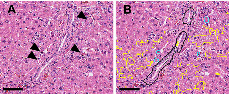
Fig. 5.
A: (Original WSI): Single cell necrosis (arrowheads) and slightly vacuolated hepatocytes were found at the periportal area (drug-induced). B: (Annotation by the algorithm): Abnormal areas (single cell necrosis and vacuolation) in Fig. 5A were annotated with light blue and yellow, respectively (Bar=100 μm).

