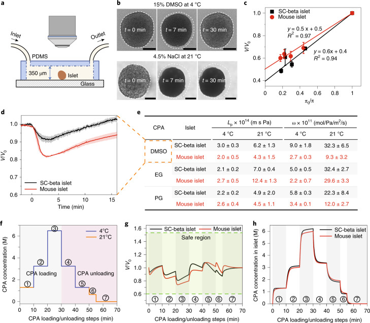
Fig. 2. Islet biophysical property measurement and CPA loading/unloading protocol design.
a, Schematic of the microfluidic device used to measure the biophysical properties of the islets (not to scale). The morphological changes of the islets were recorded via a microscope. PDMS, polydimethylsiloxane. b, Top: When subjected to 15 wt% DMSO at 4 °C, the islet first shrinks and then swells. Upon exposure to hypertonic CPA, water exits the cells, and the islet shrinks. CPA then diffuses across the cell membranes, followed by water re-entering the cell, leading to swelling back towards their initial state. Bottom: The islet remained shrunk in NaCl solution as the cells are impermeant to salt. Scale bars, 100 µm. c, Boyle–van’t Hoff plots of mouse and SC-beta islets display the normalized islet volume (V/V0) as a function of the osmolality ratio of isotonic and hypertonic NaCl solution (π0/π). The osmotic inactive volume (Vb) that does not participate in the osmotic response can be estimated by extrapolating the linear fit to π0/π = 0. Further details can be found in Methods (n = 4 for SC-beta islets, n = 8 for mouse islets). d, Normalized volume of mouse and SC-beta islets versus time demonstrating the shrink–swell behavior when exposed to 15 wt% DMSO at 21 °C (n = 3 for mouse islets, n = 9 for SC-beta islets). e, Summary table of mouse and SC-beta islets water (Lp) and CPA (ω) permeability at 4 °C and 21 °C (n = 3–9; the exact sample size can be found in Supplementary Fig. 3). The red color represents mouse islet in panels c,d,g,h, and is used in panel e to maintain consistency with the rest of the panels. f, Stepwise loading (steps 1–3) and unloading (steps 4–7) of 22 wt% EG + 22 wt% DMSO for islets. g, Modeled islet normalized volume change during CPA loading/unloading using the measured biophysical properties. The volume of both mouse and SC-beta islets remained in the safe region. h, Modeled CPA concentration in the mouse and SC-beta islets. For c–e, data are presented as mean ± s.d.

