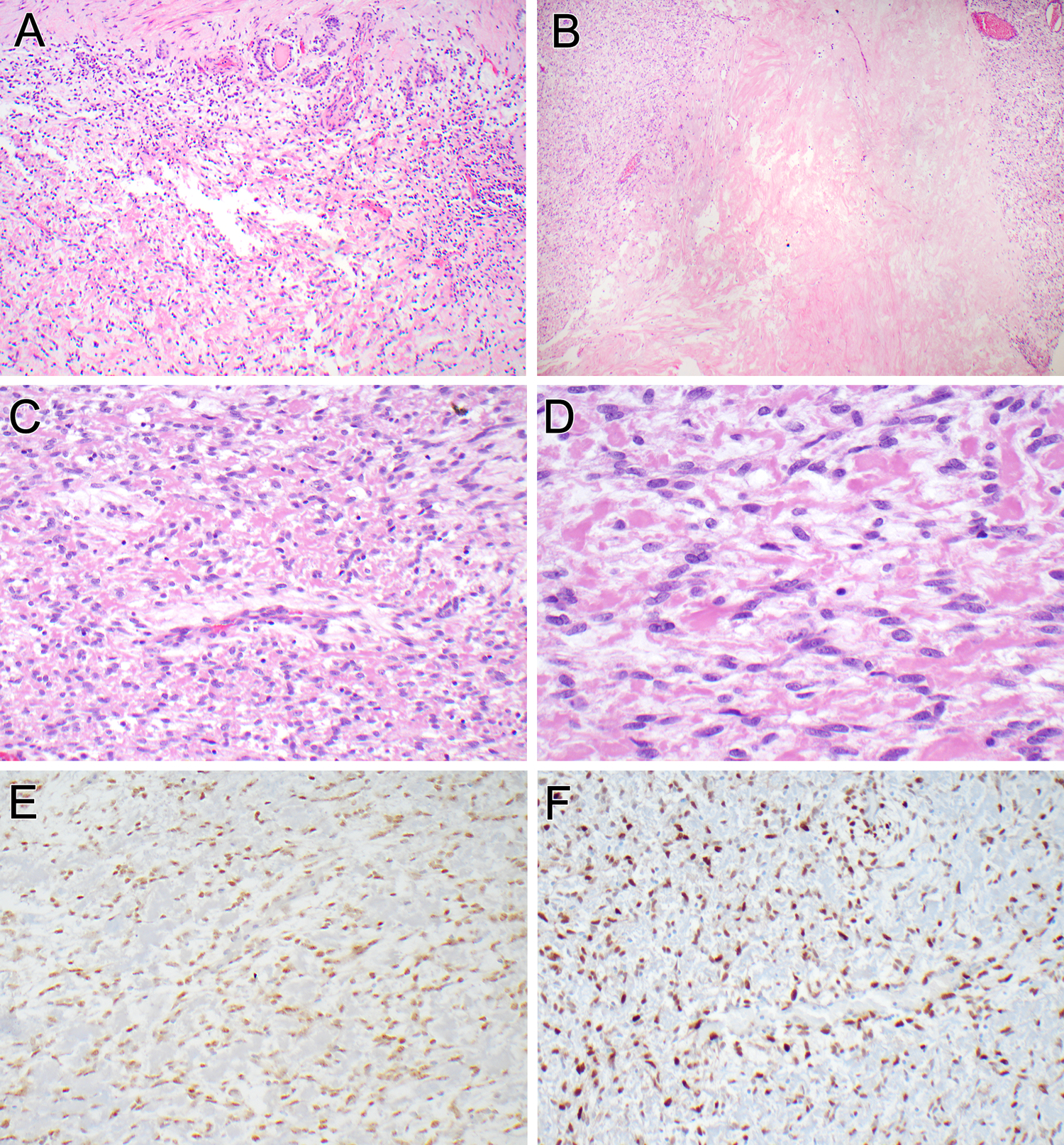
Figure 1. Pathologic findings of the renal neoplasm (case 1).

The tumor was surrounded by a variably thick fibrous capsule (top). Entrapped native renal tubules were seen embedded within the capsule and intermingled with neoplastic cells at the periphery (A). Areas of confluent, dense hyalinization were evident within the neoplasm (B). The neoplastic cells showed an ovoid to short spindle cell appearance, arranged in a perivascular distribution around intra-tumoral arterioles (C). The tumor cells were bland, with thin, short cytoplasmic extensions and ovoid nuclei with fine chromatin. The stroma separating the neoplastic cells was either myxoid or hyalinized, resembling fibrin (D). The neoplastic cells labeled diffusely for estrogen receptor (E) and cyclin D1 (E).
