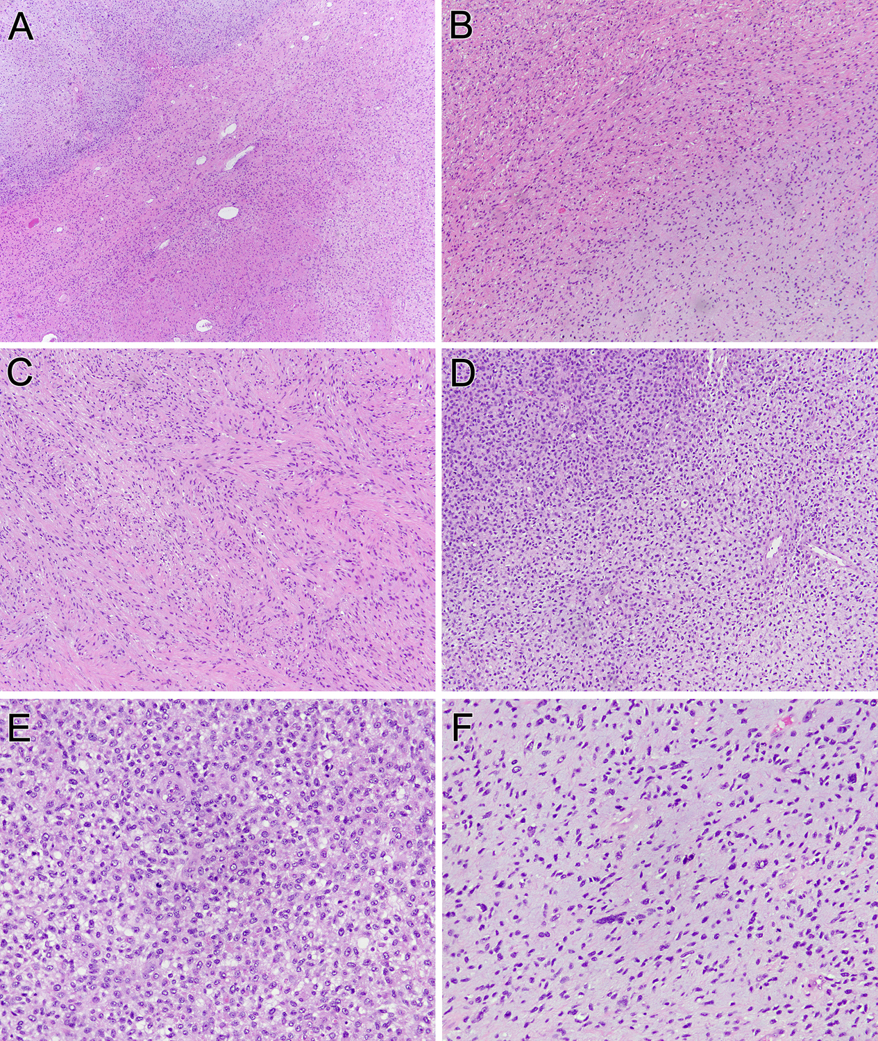
Figure 4. Pathologic features of uterine neoplasm (case 4).

This uterine neoplasm contains three components: a bland myxoid area typical of GLI1-altered neoplasms (lower right), a bland spindle cell area (middle), and a more pleomorphic myxoid area (upper left) (A). At intermediate power, one can see the transition from the bland epithelioid myxoid area (lower right) and the bland spindled area (upper left) (B). The latter component shows spindle cells with eosinophilic cytoplasm, which along with patchy desmin immunoreactivity raised the possibility of a myxoid smooth muscle neoplasm (C). The bland myxoid areas transition into more cellular areas (upper left) (D). These more overtly malignant areas demonstrate high cellularity and mitotic activity (E), as well as greater nuclear atypia (F).
