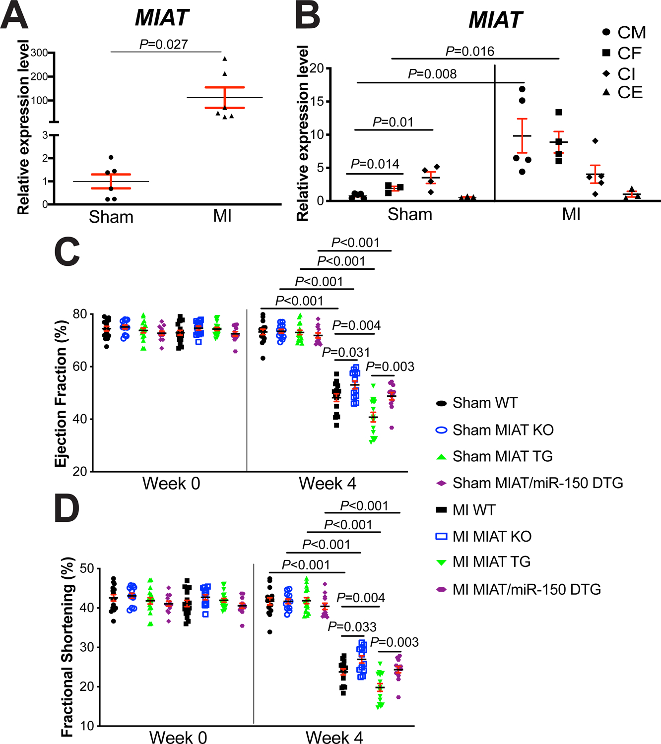
Figure 1. Myocardial Infarction-Associated Transcript (MIAT) deletion attenuates, while MIAT overexpression exacerbates cardiac dysfunction after myocardial infarction (MI) in part by repressing miR-150.

A, The expression of MIAT in left ventricles (LVs) from wild type (WT) adult mice at 4 weeks post-surgery (N=6). Data are shown as fold induction of MIAT expression normalized to glyceraldehyde-3-phosphate dehydrogenase (Gapdh). Unpaired 2-tailed t-test. B, Real-Time Quantitative Reverse Transcription (QRT)-PCR expression analysis of MIAT in cardiomyocytes (CMs), cardiac fibroblasts (CFs), cardiac inflammatory cells (CIs) and cardiac endothelial cells (CEs) isolated from adult mouse hearts at 7 days post-MI. N=3–5 per group. MIAT expression compared to Gapdh was calculated using 2−ΔΔCt, and data are shown as fold induction of MIAT expression levels normalized to CM sham. Two-way ANOVA with Tukey multiple comparison test. C-D, Transthoracic echocardiography was performed to 8 experimental groups [sham and MI of WT, MIAT knockout (KO), MIAT transgenic (TG) and MIAT/miR-150 double TG (DTG)] at week 0 and 4 post-MI. Quantification of LV ejection fraction (C) and fractional shortening (D) is shown. N=12–17 per group. Two-way repeated-measures ANOVA with Bonferroni post hoc test.
