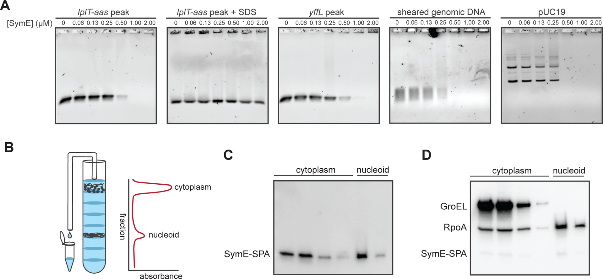
Figure 5. SymE binds DNA in vitro and is associated with the nucleoid in vivo.

(A) Electrophoretic mobility shift assays of SymE binding to 4 ng of probe corresponding to the lplT-aas ChIP peak, the yffL peak, randomly sheared E. coli genomic DNA, or purified pUC19 plasmid.
(B) Schematic of how sucrose gradient fractions were collected. Fractions containing cytoplasmic or nucleoid-associated proteins were identified by their position in the gradient and by their increased absorbances at 260 and 280 nm.
(C) Western blot of collected fractions probed with an α-FLAG antibody (which binds to the FLAG portion of the SPA tag) showing that SymE-SPA localizes to the nucleoid and cytoplasm.
(D) Same blot as in (C), but re-probed for GroEL, a chaperone, and RpoA, the alpha subunit of RNA polymerase. The bands from SymE-SPA are still visible under the RpoA bands, though the signal is weaker as RpoA and GroEL are far more abundant.
