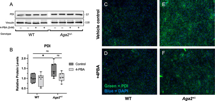
Fig. 2.

4‐PBA treatment reduces PDI levels and localization in Aga2 +/− primary osteoblast culture. (A,B) Representative Western blot and quantification of relative PDI levels in primary calvarial osteoblasts cultured and treated with 5nM 4‐PBA for 7 days. n = 7 per treatment group. A two‐way ANOVA was performed, *p < 0.05 was considered statistically significant. (C–F) Immunofluorescent images of primary osteoblasts probed with a conjugated antibody against PDI. Green = PDI, blue = DAPI, n = 6.
