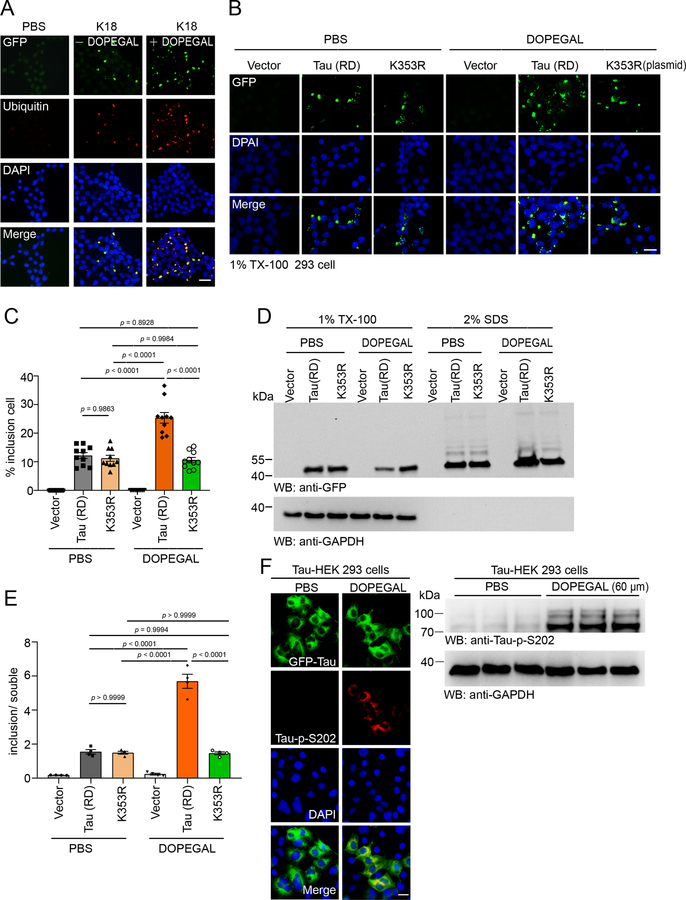
Extended Data Fig. 2. DOPEGAL promotes tau aggregation, attenuated by Tau K353R mutation.
A. Representative images showing the co-localization of aggregated tau with ubiquitin in tau PFF-treated HEK293 cells stably transfected with GFP-Tau RD in the presence or absence of DOPEGAL. Scale bar is 20 μm. B. HEK293 cells were transfected with wild-type or K353R mutant Tau RD, treated with DOPEGAL, and then transduced with tau PFFs. The green dots show tau inclusions. Scale bar is 20 μm. C. Quantification of the percentage of cells with tau inclusions. Data are shown as mean ± SEM. n = 10 per group. One-way ANOVA. D. The cells were sequentially extracted with 1% Triton X-100 lysis buffer followed by 2% SDS. Cell lysates from Triton X-100 soluble and SDS-soluble fractions were immunoblotted with GFP antibody. E. Quantification of the relative concentration of tau in the Triton X-100 soluble and SDS-soluble fractions. Data are shown as mean ± SEM. n = 4 per group. One-way ANOVA. F. The HEK293 cells stably transfected with GFP-Tau RD were exposed to DOPEGAL. The phosphorylation of tau at S202 was analyzed by immunofluorescence (red) and western blot. All data and images are representatives of three independent experiments with similar results.

