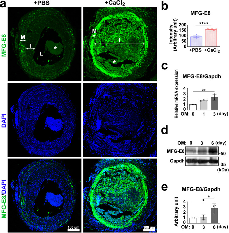
Fig. 1. Milk fat globule–epidermal growth factor 8 (MFG-E8) expression is upregulated in calcifying vessels and vascular smooth muscle cells (VSMCs).
a, b Common carotid arteries (CCAs) of wild-type mice were ligated, and phosphate-buffered saline (PBS) or CaCl2 (0.4 M) was applied on the vessels by using pluronic gel. a Representative immunofluorescence photographs display the expression of MFG-E8 in the ligated CCAs treated with PBS or CaCl2 at 21 days postligation. The neointima (I), media (M), and lumen (L) of the vessels are indicated. Blood clots in the lumen are labeled as “*”. Scale bar: 100 μm. b Quantification of fluorescence intensities of MFG-E8 in the arterial walls (PBS: nmice = 3, CaCl2, nmice = 6). Results are presented as mean ± standard error of the mean. Each data point was derived from an assessment of three sections from one individual animal. ****P < 0.0001, obtained using a t test. c–e A10 VSMCs were cultured in osteogenic medium (OM) for 6 days. c The transcript expression of MFG-E8 was evaluated through quantitative real-time polymerase chain reaction (n = 3). Data are presented as mean ± standard deviation (SD). Three independent experiments were performed. Each data point is derived from each of the three repeated experiments. **P < 0.01, obtained using one-way analysis of variance (ANOVA) followed by Tukey’s multiple comparison test. d The protein expression of MFG-E8 was evaluated through western blotting. e Quantitative analyses of MFG-E8 levels normalized to those of glyceraldehyde 3-phosphate dehydrogenase (GAPDH) were conducted (n = 3). Data are presented as mean ± SD. Three independent experiments were performed. Each data point is derived from each of the three repeated experiments. *P < 0.05, as obtained using one-way ANOVA followed by Tukey’s multiple comparisons test.

