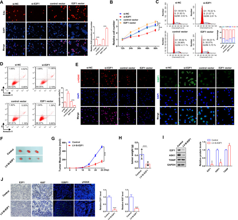
Fig. 2. E2F1 functions as an oncogene in GC.
A, B Cell proliferation assays (EdU and CCK8) were performed in MKN-45 cells transfected with si-E2F1 or E2F1 vector. C, D Representative images and histogram statistics from cell cycle and apoptosis. E Immunofluorescence staining of DNA damage markers, γH2AX and 53BP1, in MKN-45 cells transfected with si-E2F1 or E2F1 vector. F Representative images of tumors from mice implanted with control MKN-45 cells and E2F1-inhibiting MKN-45 cells. The cells were implanted subcutaneously into 6-week-old SCID mice (3 mice per group). G The time course of tumor growth. Tumor volume was measured every 5 days for 25 days after the inoculation. H The tumor weights of two groups. I Western blotting analysis of E2F1, ASK1 and TXNIP protein levels in tumors from the implanted mice. J E2F1, Ki67 and 53BP1, γH2AX staining of tumor sections obtained from two groups. All data are shown as the mean ± S.E. of three separate experiments. *P < 0.05; **P < 0.01; *** P < 0.001.

