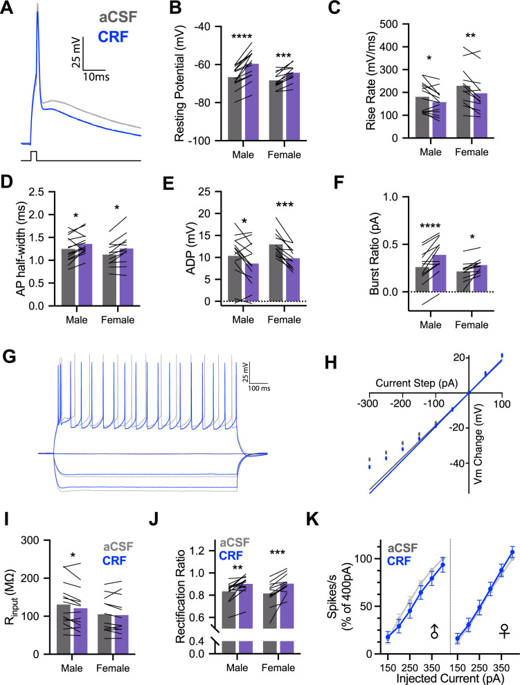
Fig. 1. CRF alters intrinsic properties of male and female insular cortex pyramidal neurons in whole-cell recordings.
A Representative single action potential (AP) recordings of deep layer insular cortex pyramidal neurons at baseline (aCSF-gray) and after application of 50 nM Corticotropin-releasing factor (CRF-blue). B CRF decreased the resting potential of male (n = 14) and female (n = 12) pyramidal neurons, FCRF(1, 24) = 72.93, P < 0.0001, with this effect being stronger in males than females as indicated by a CRF × sex interaction, FCRF × SEX(1, 24) = 5.124, P = 0.033. C Action potential rise rate was reduced by CRF in both males and females, FCRF(1, 24) = 18.93, P = 0.0002. D Action potential half-width increased following CRF application in male and female recordings, FCRF(1, 24) = 16.69, P = 0.0004. E CRF reduced the amplitude of the after depolarization (ADP) in both male and female recordings, FCRF(1, 24) = 26.83, P < 0.0001. F CRF increased the current required to trigger burst firing in male and female neurons, FCRF(1, 24) = 38.82, P < 0.0001. G Representative family of 1 s hyperpolarizing and depolarizing current injections used characterize passive membrane properties and spike rate in aCSF (gray) and after 50 nM CRF (blue). H Example steady-state current–voltage dependence plot. Input resistance was determined by linear fit and slope at 0pA and deviation from fit indicates rectification. I CRF reduced membrane input resistance in male and female neurons, FCRF(1, 24) = 5.985, P = 0.022; this effect appeared most robustly in males. J CRF increased rectification of membrane potential in males and females, FCRF(1, 24) = 35.28, P < 0.0001. K CRF did not alter firing rates in response to 1 s depolarizing current injections in either males or females. Bar graphs indicate mean with individual replicates, line graphs mean (±SEM). *P < 0.05, **P < 0.01, ***P < 0.001, ****P < 0.0001 (Sidak’s tests).

