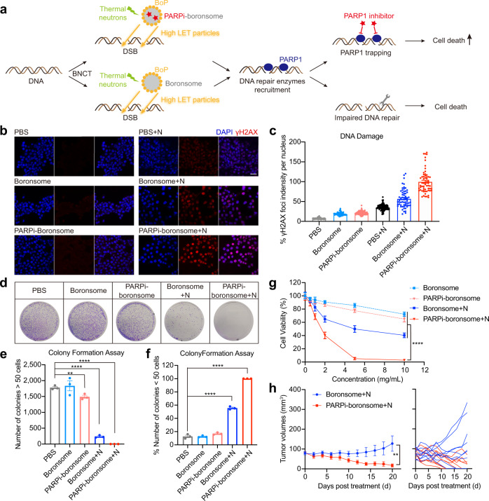
Fig. 6. DNA damages repair was significantly inhibited by BNCT with PARP1 inhibitor–loaded boronsome.
a Schematic illustration of the synergy between PARP1 inhibition and BNCT. b Representative immunofluorescence images were recorded using confocal microscopy. Nuclei were stained with DAPI (blue) and antibody to γH2AX (red). Scale bar, 50 μm. c Quantification relative γH2AX red fluorescence in densities per nucleus. Each dots represents the data in a nucleus shown in b. PBS, 124 cells; Boronsome, 79 cells; PARPi-boronsome, 167 cells; PBS + N, 117 cells; Boronsome + N, 61 cells; PARPi-boronsome + N, 60 cells. Three independent experiments were performed and representative results are shown. d–f Colony formation assay of 4T1 cells cultured in 10-cm dishes treated with boronsome, PARPi-boronsome, boronsome+neutron (N) and PARPi-boronsome+neutron (N). d Colonies were stained with crystal violet. e Quantification of the number of colonies consisting of at least 50 cells (n = 3, two-tailed unpaired Student’s t test, **p = 0.0087, ****p < 0.0001). f Percentage of the number of colonies <50 cells (n = 3, two-tailed unpaired Student’s t test, ****p < 0.0001). g Cell viability of 4T1 cells under various conditions, detected by CCK-8 assay 24 h post treatment (n = 6, two-tailed unpaired Student’s t test for data of 10 mg/kg, ****p < 0.0001). h Average tumour volumes (n = 9) of each group of mice (two-tailed paired Student’s t test, ** p = 0.0025). Source data are provided as a Source Data file.

