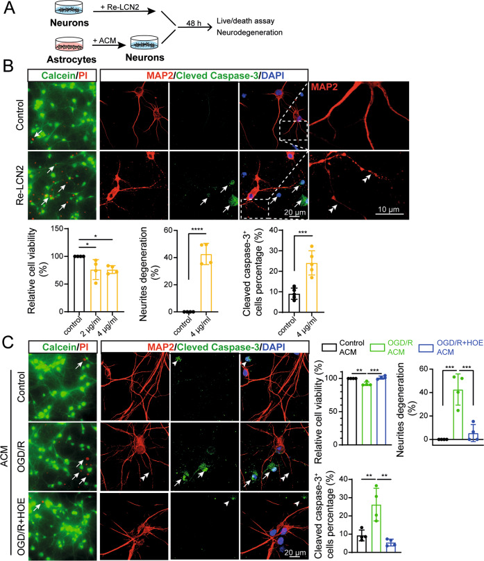
Fig. 5. ACM from NHE1 inhibited astrocytes induced by in vitro ischemia are protective against neuronal degeneration.
A Experimental protocol. Cartoon illustrates primary neurons treated with recombinant LCN2 (Re-LCN2) and ACM collected from different treatment groups at 24 h REOX. B Left panel: Representative Calcein-AM (green)/propidium iodide (red) staining of primary neurons treated with Re-LCN2 for 48 h. Right Panel: Representative confocal images of MAP2 and cleaved caspase-3 stained neurons treated with 4 μg/ml Re-LCN2 for 48 h. Arrows: high expression. Arrowheads: low expression. Double arrowheads: neurites degeneration. Summary data shows quantification of cell viability, percentage of neurite degeneration and cleaved caspase-3 expression. Data are mean ± SD, n = 4. *p < 0.05; **p < 0.01; ***p < 0.001; ****p < 0.0001 via one-way ANOVA and unpaired t-test. C Representative Calcein/PI staining and MAP2/cleaved caspase-3 staining of primary neurons treated with ACM of control, OGD/R and OGD/R + HOE642 treated astrocytes for 48 h. Arrows: high expression. Arrowheads: low expression. Double arrowheads: neurites degeneration. Summary data shows quantification of cell viability, percentage of neurite degeneration and cleaved caspase-3 expression. Data are mean ± SD, n = 4. *p < 0.05; **p < 0.01; ***p < 0.001 via one-way ANOVA and unpaired t-test.

