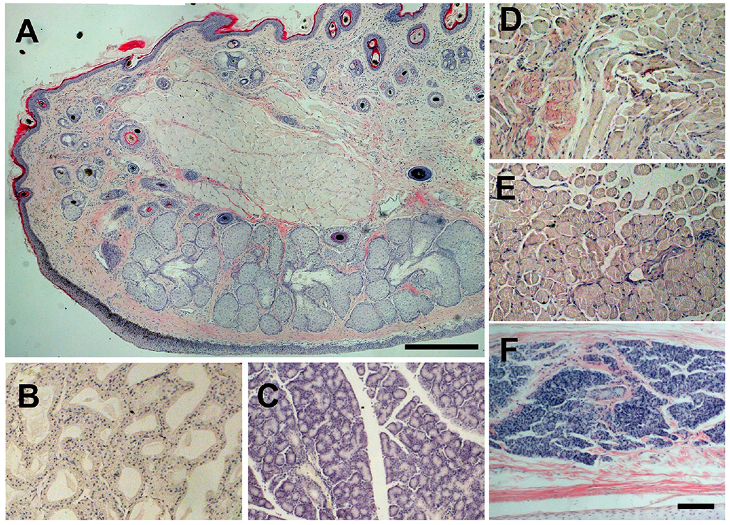
Figure 4.

Histology of adnexal tissues after SMG injection. SMG at 40mM was injected into the sT and the eye fixed at 5 days after the injection. Paraffin sections of adnexal tissues with H/E staining are shown. A, eyelid, B, Harderian gland, C, lacrimal gland, D, superior oblique muscle, E, superior rectus muscle, F, nictitating membrane, Bar, 200 μm in panel A, 100 μm in all other panels.
