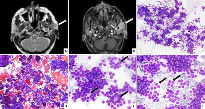
Fig. 1.
Axial T2 weighted (A) and post gadolinium fat saturated T1 weighted (B) images of contrast-enhanced MRI show an irregular solid lesion with cystic components in left parotid gland (arrow) infiltrating into the masseter and abutting the mandibular ramus, showing hyperintense signal on T2 weighted images and intense contrast enhancement. FNAC shows a tumor with predominantly trabecular (C; HE, 100× ) and focal pseudopapillary architecture (D; HE, 200× ). The cells form rosette-like structures (arrows) at places (E; HE, 400× ). They appear monotonous, have scant granular cytoplasm and round to ovoid nuclei, some of which are eccentrically placed, imparting a plasmacytoid (arrows) appearance (F; HE, 400× )

