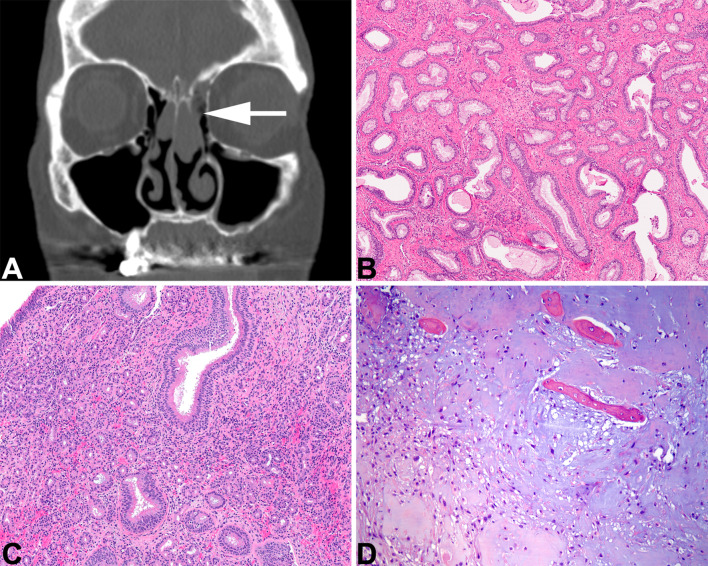
Fig. 1.
Sinonasal hamartomas. A Respiratory epithelial adenomatoid hamartoma. An axial computed tomography shows expanded olfactory cleft (white arrow) with a soft tissue-density mass. B Evenly spaced glandular units have a prominent basement membrane surrounding them. C. Seromucinous hamartoma shows a proliferation of small eosinophilic glands in the stroma, lacking destructive growth. D A chondromesenchymal hamartoma shows cartilaginous nodules with a myxoid stroma and islands of bony tissue (courtesy Dr. D. Baumhoer)

