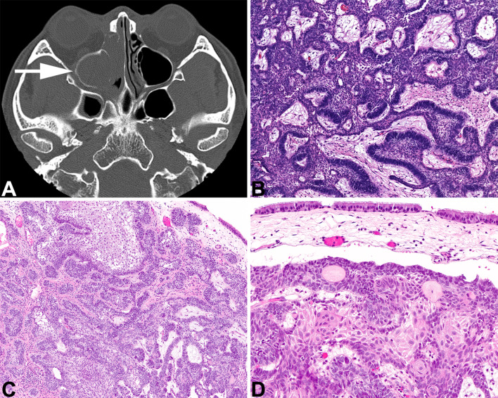
Fig. 9.
Sinonasal ameloblastoma. A An axial CT shows an ethmoid sinus mass expanding and filling the sinus with a soft tissue-density mass. B A central stellate reticulum with ameloblastic palisade. C The surface respiratory epithelium overlies the proliferation of ameloblastoma. D An intact, ciliated respiratory-type epithelium is seen overlying a proliferation of columnar, basaloid cells associated with a well-developed central stellate reticulum

