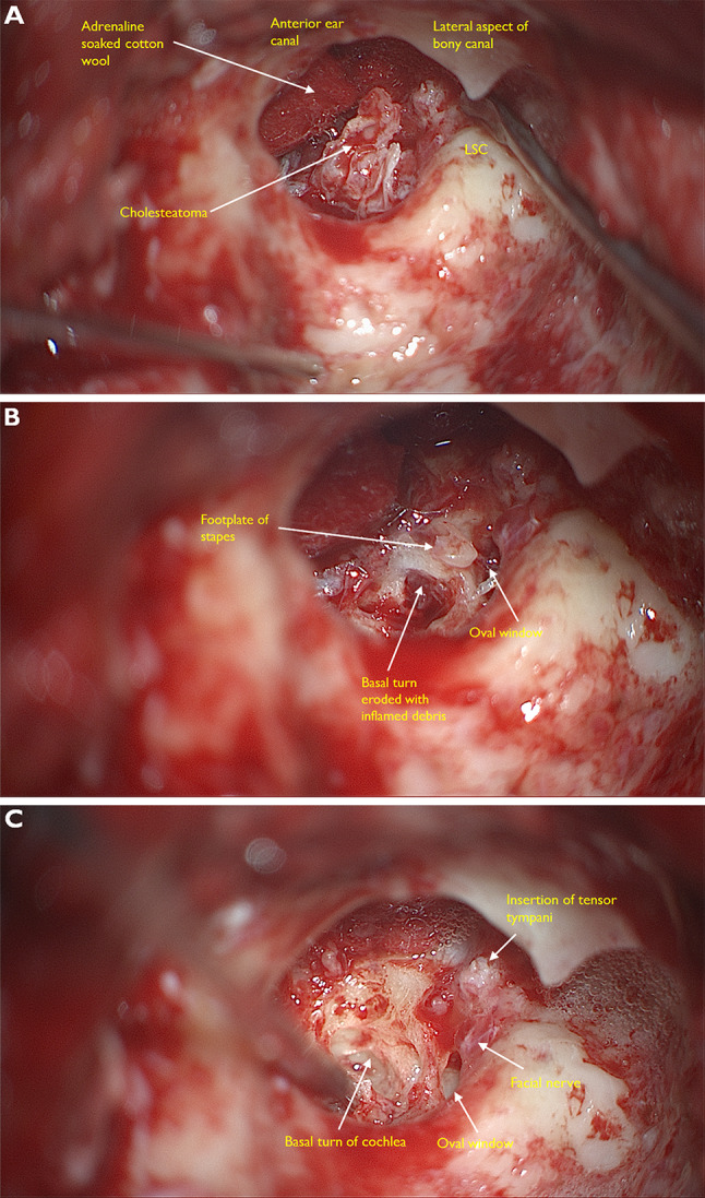
Fig. 5.

Intraoperative microscopic views taken at mastoidectomy for cholesteatoma*. A The ear canal has been taken down and we can see into the middle ear. The cholesteatoma appears as a friable white mass. B This image shows erosion of the footplate of the stapes. C On further dissection the anatomy of the cochlear is clearly visible. Round window is totally eroded and there is complete destruction of the cochlea promontory. The tympanic segment of the facial nerve is usually in a bony canal which can sometimes be dehiscent (~ 10% of patients with healthy ears). Here it is exposed, possibly inflamed and damaged due to irritation from the overlying cholesteatoma. *Images courtesy Mr Harry Powell Consultant ENT surgeon & Dr Charlotte Arnold (trainee ENT surgeon) Guys & St Thomas’s NHS Foundation trust London UK
