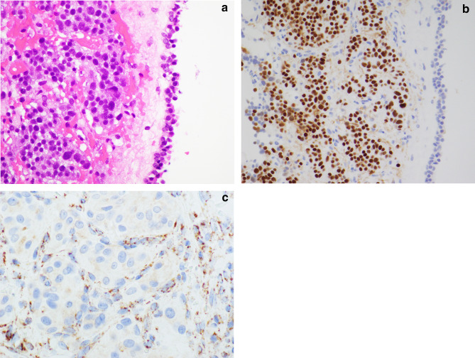
Fig. 5.
Middle ear paraganglioma: SDHx-related paragangliomas from the head and neck usually have small cells with clear cytoplasm (a). The tumor cells express markers of neuroendocrine differentiation, including insulinoma associated protein1/INSM1 (b), synaptophysin, and chromogranin A, as well as nuclear GATA3 and cytoplasmic catecholamine-synthesizing enzymes, tyrosine hydroxylase and dopamine β-hydroxylase. SDHB-immunohistochemistry is negative in tumour cells of SDHx-related tumors (c), and only capillary endothelial cells are positive as internal controls. On the other hand, tumour cells in paragangliomas with non-SDH-mutation demonstrate numerous immunoreactive granules in the cytoplasm

