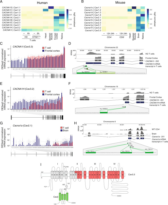
Fig. 7. Lack of full-length transcripts of VGCC α1 subunits in T cells.
A, B Absolute mRNA expression levels of α1 subunits of VGCCs in human (A) and mouse (B) T cells and reference tissues. A CD4+ T cells were left unstimulated (−) or stimulated for 6 h with anti-CD3 + CD28 antibodies. HD represents the averaged data from three individual healthy donors (HD) and a patient with a STIM1 null mutation (c.497 + 776 A > G; STIM1null). B Mouse CD4+ and CD8+ T cells were left unstimulated (−) or stimulated for 12 or 24 h with anti-CD3 + CD28 antibodies. VGCC expression in T cells in (A, B) was analyzed by RNA sequencing and compared to that in human and mouse heart, skeletal muscle, frontal cortex, whole brain and retina using published datasets (Supplementary Table 2). C–F Exon usage of human CACNA1I (Cav3.3) (in C, D) and CACNA1H (Cav3.2) (in E, F) in CD4+ T cells and frontal cortex. C, E Normalized mRNA expression in unstimulated T cells from n = 3 HDs (averaged; red) and frontal cortex (blue) per exon. D, F Transcript levels (as RPKM, reads per kb of transcript, per million mapped reads) in frontal cortex and CD4+ T cells from an individual HD superimposed on exons 12–15 (for CACNA1I in D) and exons 12–14 (for CACNA1H in F). In T cells, exon 12 and exon 13 are the first transcribed exons of CACNA1I and CACNA1H, respectively. Green boxes and nucleotide sequences indicate predicted alternative transcriptional start sites (TSS). G Exon usage of Cacna1a (Cav2.1) in mouse CD4+ T cells (red) and brain (blue). Normalized Cacna1a mRNA expression per exon and exon-intron structure. H Cacna1a transcript levels (as RPKM) in brain and CD4+ T cells from mice superimposed on exons 32–36. In T cells, exon 34 is the first transcribed exon. Green boxes and nucleotide sequences indicate predicted alternative TSS. I Membrane topology model of the predicted N-terminally truncated Cav3.3 protein in T cells lacking channel domain I and part of domain II. AID α1 interacting domain, ABP α1 binding pocket.

