
Figure 5. ACLY controls acetyl-CoA production and Th17 cell effector function.
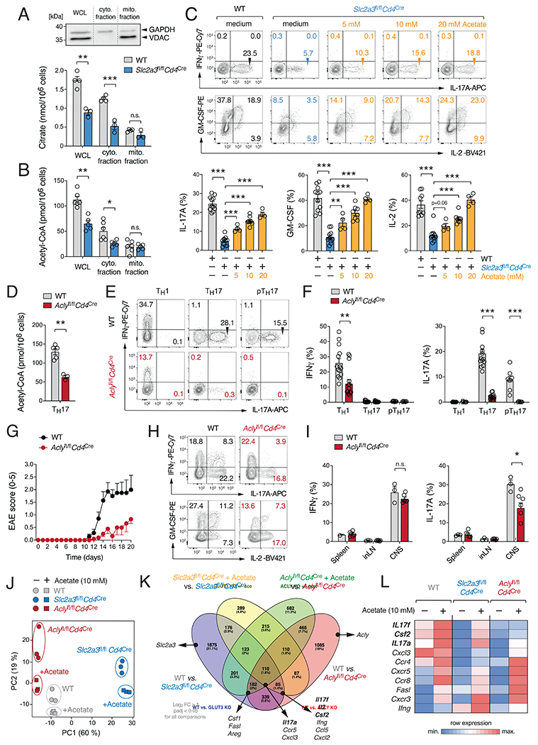
(A and B) Analysis of subcellular citrate (A) and acetyl-CoA (B) in whole cell lysates (WCL) and isolated cytosolic and mitochondrial fractions; means ± SEM of 5-6 mice. Immunoblot analysis of cytosolic (GAPDH) and mitochondrial (VDAC) proteins. (C) Exogenous acetate rescues impaired cytokine production of GLUT3-deficient Th17 cells. Flow cytometric analyses of IFNγ, IL-17, IL-2 and GM-CSF expression of WT and Slc2a3fl/flCd4Cre Th17 cells treated with acetate; means ± SEM of 4-11 mice. (D-I) ACLY controls Th17 cell effector function. (D) Quantification of acetyl-CoA in WCL of WT and Aclyfl/flCd4Cre Th17 cells; means ± SEM of 3-5 mice. (E and F) Flow cytometric analysis of IFNγ and IL-17 production by WT and ACLY-deficient (Aclyfl/flCd4Cre) T cells; means ± SEM of 6-14 mice. (G–I) Inactivation of ACLY in T cells prevents autoimmune encephalomyelitis. (G) Clinical EAE scores of WT and Aclyfl/flCd4Cre mice after MOG35-55 immunization; means ± SEM of 7-10 mice per cohort. (H and I) Flow cytometric analyses of IFNγ and IL-17 production of CD4+ T cells from WT and Aclyfl/flCd4Cre mice; means ± SEM of 3-6 mice. (J-L) Global gene expression analysis of WT, GLUT3-deficient (Slc2a3fl/flCd4Cre) and ACLY-deficient (Aclyfl/flCd4Cre) Th17 cells in the presence and absence of acetate. (J) Principal component (PC) analysis of WT, GLUT3-deficient and ACLY-deficient Th17 cell gene expression data. (K) Venn diagram analyses comparing GLUT3-deficient, ACLY-deficient and WT Th17 cells in the presence or absence of 10 mM acetate; > 2-fold expression change and padj < 0.05. (L) Heatmap analysis of selected gene expression in WT, GLUT3- and ACLY-deficient Th17 cells. *, p<0.05; **, p<0.01, ***, p<0.001 by unpaired Student’s t-test (A-D), (F) and (I).