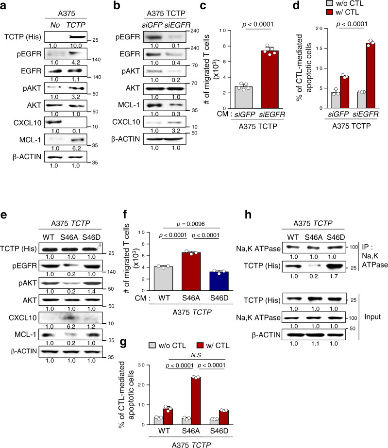
Fig. 5. TCTP phosphorylation is crucial to activating the EGFR/AKT signaling pathway via binding with Na, K ATPase.
a The protein levels of TCTP, EGFR, pEGFR, pAKT, AKT, MCL-1, and CXCL10 were measured by Western blot analysis. b The protein levels of pEGFR, EGFR, pAKT, AKT, MCL-1, and CXCL10 in SiGFP- or siEGFR-treated A375 TCTP cells were analyzed by Western blots. c SiGFP- or siEGFR-treated A375 TCTP cells CM was added to the lower chamber, and CD8+ T cells were plated in the upper chamber. The T cells migrated into the lower chamber media were collected after 6 h and counted. d CFSE-labeled tumor cells were exposed to NY-ESO1-specific CTLs and the frequency of CFSE + apoptotic tumor cells was determined by flow cytometric analysis of active-caspase-3. e–h A375 cells were transfected with FLAG-TCTP wild type (TCTP), FLAG-TCTP S46A mutant, or FLAG-TCTP S46D mutant. e Activation of EGFR, AKT signaling and expression of MCL-1 and CXCL10 were analyzed by western blot assays. f T cell chemotaxis assays were performed by using the indicated tumor cell-derived CM. g Active-caspase-3+ apoptotic tumor cells were analyzed by flow cytometry after incubation with CTLs. h Cell lysates were immunoprecipitated with anti-Na, K ATPase antibody. The immunoprecipitated proteins were analyzed by Western blot assays. The data are representative of three separate experiments. The numbers below the blot images indicate the expression as measured fold change a, b, e, and h. The p values by two-tailed t test c, two-way ANOVA d, g, and one-way ANOVA f are indicated. N.S, not significant. The error bars represent mean ± SD. Source data are provided as a Source data file.

