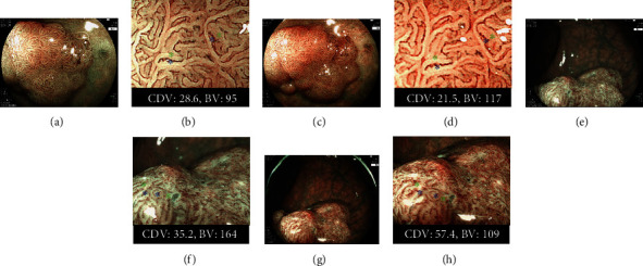
Figure 3.

The comparison of Color difference value (CDV) and bright value (BV) between the BLI-LED and BLI-LASER endoscopic systems. (a). A nonpolypoid of 20 mm on the rectum. Histopathology: high-grade adenoma. BLI-LED: JNET Type 2B. (b) The CDV and BV for two points between the vessels (the red signs) and surrounding whitish area (the white signs) with BLI-LED were 28.6 and 95. (c) BLI-LASER of the same tumor: JNET Type 2A. (d) The CDV and BV were 21.5 and 117. (e) A nonpolypoid of 25 mm on the rectum. Histopathology: T1b cancer. BLI of LED: JNET Type 3. (f) The CDV and BV were 35.2 and 164. (g) BLI-LASER of the same tumor: JNET Type 3. (h) The CDV and BV were 57.4 and 109.
