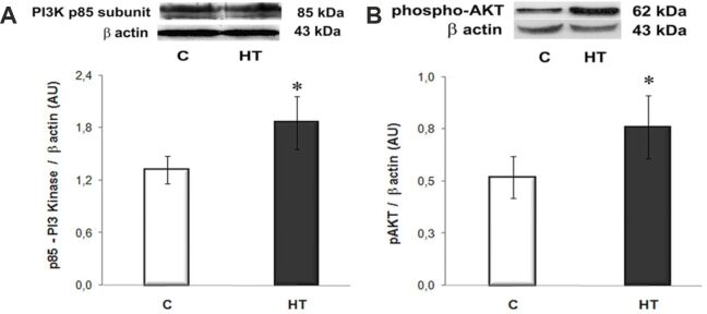
Figure 3.
The expression levels of PI3K (p85 subunit) (A) and phospho-AKT (Ser473) (B) are significantly increased in the LVs of HT hamsters compared to controls [C] and assessed by Western blot. Representative immunoblots and the densitometric analysis (AU) are displayed in the upper and lower panels, respectively. β-Actin was used as protein loading control. n: 3–4 hamsters in each group. *p<0.05 vs. [C] group. AKT: Protein kinase B; AU: Arbitrary units; HT: Hypertensive; LV: Left ventricle; PI3K: Phosphoinositide 3-kinase

