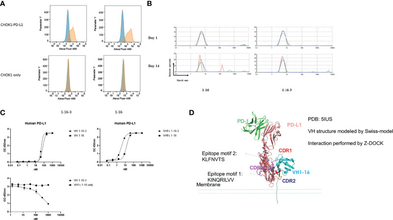
Figure 3.
Characterization of VH domains 1-16 and 1-16-3. (A) Flow cytometry analysis for binding of VH 1-16 and 1-16-3 to PD-L1 expressed on the surface of CHOK1 cells; blue peaks represent cells stained with AF488 conjugated secondary antibody only, red peaks represent cells stained with isotype control and then AF488 conjugated secondary antibody, yellow peaks represent cells stained with VH domains and then AF488 conjugated secondary antibody; (B) Dynamic light scattering analysis for evaluation of the aggregation propensity of domain s domains 1-16 and 1-16-3; Curves with different color are different repeats; (C) ELISA of 1-16 and 1-16-3 against PD-L1 with or without human IgG1 Fc, and their competition; (D) Structural modelling of PD-L1 with VH 1-16. Epitopes were labeled in the figure.

