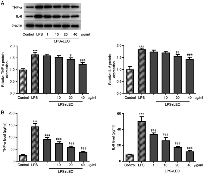
Figure 3.
LEO attenuates the LPS-induced inflammatory response in BEAS-2B cells. (A) Western blot analysis was used to determine the protein expression levels of TNF-α and IL-6. (B) ELISA was used to quantify the concentrations of TNF-α and IL-6. Data are presented as the mean ± standard deviation of three independent experiments. ***P<0.001 vs. control; #P<0.05, ##P<0.01, ###P<0.001 vs. LPS. LEO, leonurine; LPS, lipopolysaccharide.

