Figure 3.

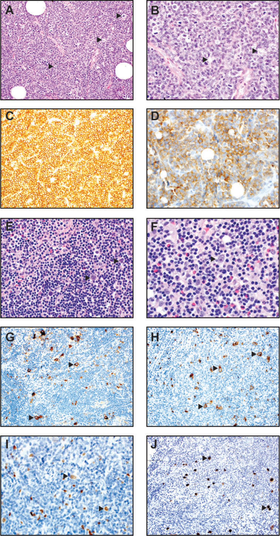
Burkitt lymphoma (A-D) and classic Hodgkin lymphoma, mixed cellular type (E-J). (A) At medium power of H&E (200x), a characteristic starry-sky pattern is present. The “stars” (arrowheads) are represented by tingible-body macrophages and the “sky” is composed of neoplastic lymphoid cells. (B) At high magnification of H&E slide (400x), the neoplastic cells are intermediate-sized, with slightly irregular nuclear contours, few nucleoli, and basophilic cytoplasm. Arrowheads mark tingible-body macrophages. Many mitotic figures and apoptotic debris are also present. (C) The neoplastic cells are strongly positive for the B cell marker CD20 (200x). (D) They are strongly positive for CD10 (400x) and BCL6 (not shown) consistent with a germinal center phenotype. (E) In this lymph node, the high-power magnification of H&E (400x) shows binucleate Reed-Sternberg (RS) cells (highlighted by arrowheads) and mononucleate Hodgkin (H) cells within a polymorphous background composed of small lymphocytes, histiocytes, eosinophils, and plasma cells. (F) A higher power magnification of H&E (600x) shows similar morphologic features, with an arrow pointing to a binucleate Reed-Sternberg H&E (600x). (G) Immunohistochemistry for CD30 (200x) highlights scattered RS and H cells (arrowheads). (H) Immunohistochemistry for CD15 (200x) highlights scattered RS and H cells (arrowheads). (I) The neoplastic RS and H cells (arrowheads) demonstrate variably weak positivity for PAX-5 immunostain. (J) The neoplastic RS and H cells (arrowheads) in this case are positive for EBV-encoded small RNA by in situ hybridization (400x).
