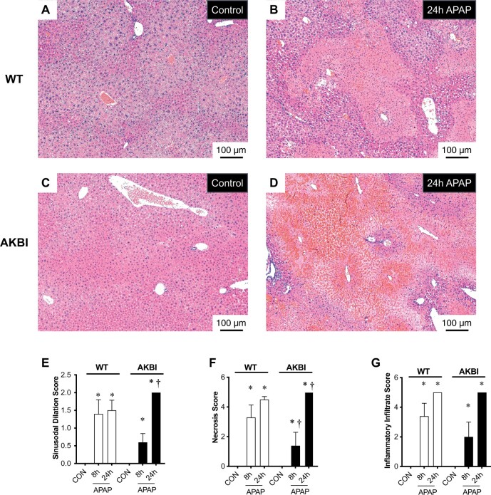
Figure 3.
Time course of acetaminophen (APAP)-induced hepatic injury in wild type (WT) and AKBI mice. WT and AKBI (IkappaB beta knock-in mice) were exposed to APAP (280 mg/kg, IP) for 0 or 24 h. A–D, Representative hematoxylin and eosin (H&E)-stained hepatic sections from WT (A and B) and AKBI (C and D) control and APAP exposed (24 h). Internal scale bar 100 μm. E–G, Blind histopathologic evaluation of H&E-stained hepatic sections scored for E, necrosis, F, inflammatory infiltration, and G, sinusoidal dilatation. White columns represent WT mice and black columns represent AKBI mice (n = 4–6). Data are expressed as mean ± SEM; *p < .05 versus unexposed genotype-matched control; †p < .05 versus similarly time-matched APAP-exposed WT.

