FIGURE 2.

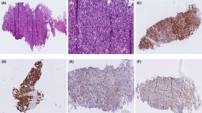
Histology examination (H&E) high‐power magnified image ×20 (A) and ×40 (B) of the tumor. Immunohistochemical findings include positive Melan‐A (C), SOX‐10 (D), HMB‐45 (E) and focally positive S‐ 100 (F)

Histology examination (H&E) high‐power magnified image ×20 (A) and ×40 (B) of the tumor. Immunohistochemical findings include positive Melan‐A (C), SOX‐10 (D), HMB‐45 (E) and focally positive S‐ 100 (F)