Figure 1.
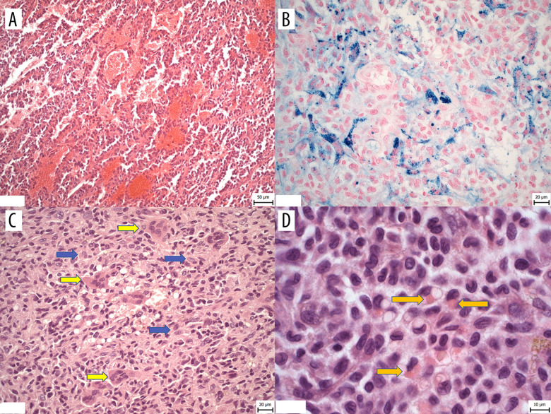
Representative histopathology images. (A) The vasoformative and reticular growth patterns of tumor with vascular-like spaces filled with erythrocytes. (B) Deposits of hemosiderin pigment in siderophages in fibrous neoplastic stroma of tumor (iron Perls staining). (C) Solid growth patterns of tumor consisting of neoplastic cells with occasional larger cells of round-to-oval shape, with abundant eosinophilic cytoplasm (marked by blue arrows) and osteoclast-like cells (marked by yellow arrows) (HE, magnification 40×). (D) Higher magnification of neoplastic cells showing round-to-oval nuclei with irregular folds and hemophagocytosis in occasional neoplastic cells (indicated by arrows) (HE, magnification 100×).

