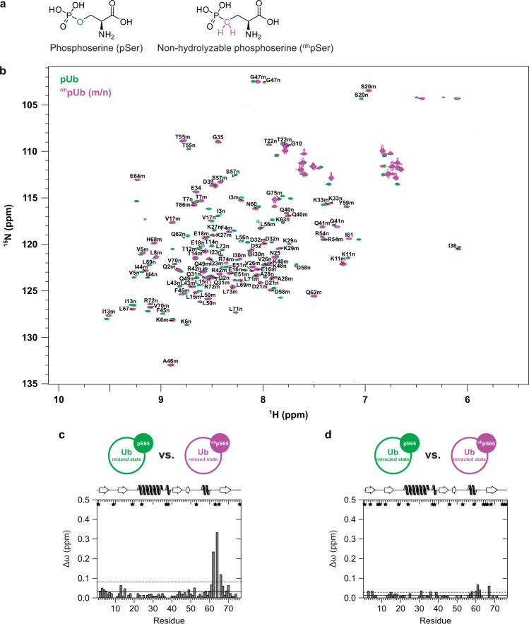
Fig. 3. NMR spectroscopic characterization of nhpUb.
a Structure of l-O-phosphoserine (pSer) and its non-hydrolyzable analog l-2-Amino-4-phosphonobutyric acid (nhpSer). b Two-dimensional heteronuclear 1H–15N HSQC NMR spectra of pUb (colored in green) and nhpUb (colored in purple) are superimposed. Assigned backbone amide resonance signals from the relaxed state (m) and the retracted state (n) of nhpUb are labeled in the spectrum by using the one letter code for amino acids. c, d Weighted chemical shift perturbation (CSP, Δω) mappings comparing either the relaxed states (c) or the retracted states of pUb and nhpUb with each other (d). Δω values exceeding the horizontal straight line are larger than the mean and Δω values exceeding the horizontal dashed line are larger than the mean plus one standard deviation. Residues that could not be assigned in both Ub species are indicated by an asterisk. Secondary structure elements according to the NMR solution structure of Ub (PDB ID 1D3Z)74 are depicted on top of the graphs.

