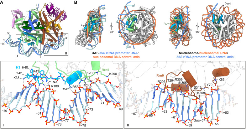
Fig. 2. Interactions of UAF with promoter DNA.
(A) Overview of the UAF-DNA interface. Insets show the chemical environments of the Rrn5-H3-contact site (I) and Rrn9 contact site (II), respectively. Participating secondary structure elements are colored. Bases in the promoter sequence relative to the 35S rRNA transcription start site are indicated. (B) 35S rRNA promoter DNA position relative to the nucleosome. UAF is aligned to the yeast nucleosome (PDB, 1ID3) (26) based on Rrn5 (green) and the proximal H3 (blue). Left: UAF-TBP-DNA superposed with the central axis of nucleosomal DNA (orange), calculated with Curves+ (51). Right: Corresponding views of the nucleosome. Superposed is the central axis of UAF-bound DNA (dark blue). The H3 (cyan) and H4 (green) subunits used for alignment with UAF are colored, and the dyad axis is indicated.

