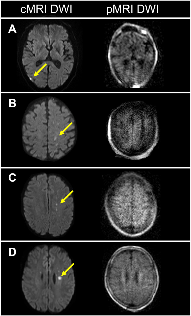
Fig. 2. Patients with ischemic stroke with infarcts not visualized by pMRI.
Four patients with ischemic stroke (A to D) had foci of restricted diffusion (yellow arrows) that were detected solely by conventional MRI (cMRI) (1.5/3 T) DWI sequences, but not pMRI DWI. (A) 7-mm-diameter infarct. (B) 10-mm infarct. (C) 4-mm infarct. (D) 7-mm infarct.

