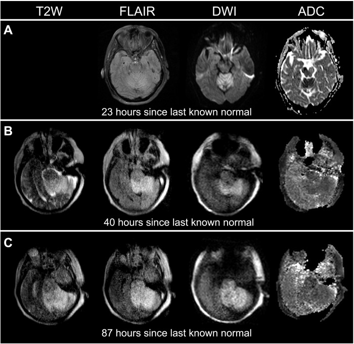
Fig. 4. Serial imaging with pMRI facilitated dynamic monitoring of acute ischemic stroke progression in a 66-year-old female with a brainstem stroke.
(A) The SOC MRI (3 T) was obtained 23 hours since last known normal. (B) The first pMRI exam was obtained 40 hours since last known normal. (C) The second pMRI exam was obtained 87 hours since last known normal. Serial pMRI exams (B and C) revealed an evolution and extension of cerebral infarction from the cerebellum into the brainstem (70.430 to 97.681 cm3). The SOC MRI exam did not include a T2W sequence.

