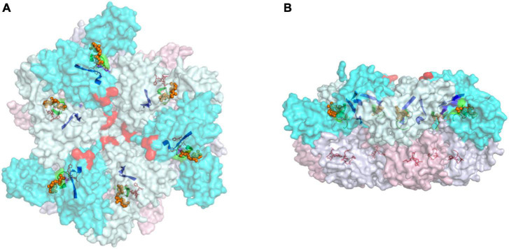
FIGURE 2.
Top (A) and side (B) views of the cytoplasmic region of hexameric FtsH complex from T. thermophilus (PDB:2DHR). Annotations: protomers are defined according to the status of its ATPase domains (“open” confirmation in cyan and “closed” confirmation in pale-cyan). The protease domains of “open” protomers are labeled pink, and the “closed” counterparts are in blue-white. Selected motifs presented in a non-translucent color include Walker A (green ribbon) and B (blue ribbon), and the zinc coordinates of the protease site (red sticks). The proposed FVG motif responsible for substrate binding is displayed as a red sphere, and ADP molecules are shown as orange spheres.

