. 2022 Apr 20;11:e72599. doi: 10.7554/eLife.72599

Figure 3. Expanded expression of olfactory co-receptors.

(A–D) Comparing knock-in innervation patterns of the antennal lobe (AL) with what has previously been reported for each co-receptor. Co-labeling experiments with each co-receptor knock-in line driving QUAS-GFP (green) and the corresponding transgenic co-receptor Gal4 line driving UAS-mCD8::RFP (anti-CD8, orange). The nc82 antibody labels synapses (magenta) and is used as a brain counterstain in these and all subsequent brain images. (A) The Orco-T2A-QF2 knock-in labels more glomeruli than the Orco-Gal4 line. Top: maximum intensity projection of full z-stack showing two additional glomeruli labeled by the knock-in, VM4 (Ir8a+/Ir76b+/Ir25a+) and VL2a (Ir8a+). Middle: subset of z-stack with a box around the V glomerulus. Bottom: zoom of boxed region showing sparse innervation of the V glomerulus (Gr21a+/Gr63a+) by the knock-in but not the Gal4 line. Asterisk indicates antennal nerve that is outside the V glomerulus. In the sub z-stack and zoom panel, gain has been increased in the GFP channel to visualize weak labeling more clearly. (B) The Ir8a-T2A-QF2 knock-in also drives GFP expression in more glomeruli than previously reported, including the outlined VL1 glomerulus (Ir25a+). (C) In the brain, Ir76b-T2A-QF2>GFP olfactory neurons innervate the ALs, while gustatory neurons from the labella innervate the subesophageal zone (SEZ, arrowhead). Top: both the Ir76b knock-in and transgenic Gal4 line label more glomeruli than previously reported, including VL1 (Ir25a+) and DP1l (Ir8a+). Bottom: the Ir76b-T2A-QF2 knock-in labels several Orco+ glomeruli, such as DC3 and VC4 (outlined). In the subset, gain has been increased in the GFP channel to visualize weakly labeled glomeruli more clearly. (D) The Ir25a-T2A-QF2 knock-in drives GFP expression broadly in the antennal lobes and SEZ (arrowhead). Ir25a+ neurons innervate many Orco+ glomeruli, such as those outlined. The transgenic Ir25a-Gal4 line labels a subset of the knock-in expression pattern. N = 3–10 for co-labeling experiments, N = 5–15 for additional analyses of the knock-in lines alone. Scale bars = 25 µm, except zoom panel scale bar = 10 µm. See also Figure 3—figure supplements 1 and 2, Table 3, and Figure 3—source data 1 and Figure 3—source data 2.

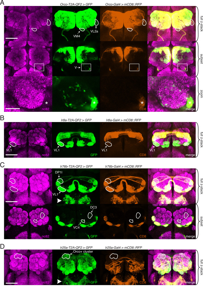
Figure 3—source data 1. Knock-in antennal lobe analyses.
Figure 3—source data 2. Examples of new glomerular expression in knock-in lines.

Figure 3.

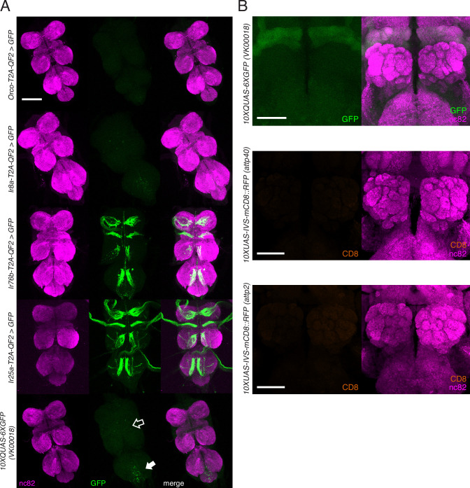
Figure 3—figure supplement 1. Knock-in expression in the adult ventral nerve cord (VNC) and reporter expression in the brain.

Figure 3—figure supplement 1.

(A) In adult flies, two of the four knock-ins (Ir76b-T2A-QF2, third row, and Ir25a-T2A-QF2, fourth row) drive GFP expression in neurons innervating the VNC. This likely reflects the role of these co-receptors in gustation. There is weak GFP expression in the reporter control (bottom row) in the abdominal neuromere (filled arrow), as well as the accessory mesothoracic neuropil (empty arrow), which can be seen in the two other knock-ins (Orco-T2A-QF2, first row, and Ir8a-T2A-QF2, second row). This nonspecific expression is likely from the reporter itself rather than driven by the two knock-ins. (B) Control brains showing the QUAS-GFP reporter alone (top) and the UAS-mCD8::RFP reporters alone (middle and bottom). These reporters were used for all brain images in Figures 35. The QUAS reporter weakly labels the mushroom bodies. The VNC from the fly in (A, fourth row) is also used in Figure 2—figure supplement 1E. Scale bars: 100 µm in (A), 50 µm in (B).
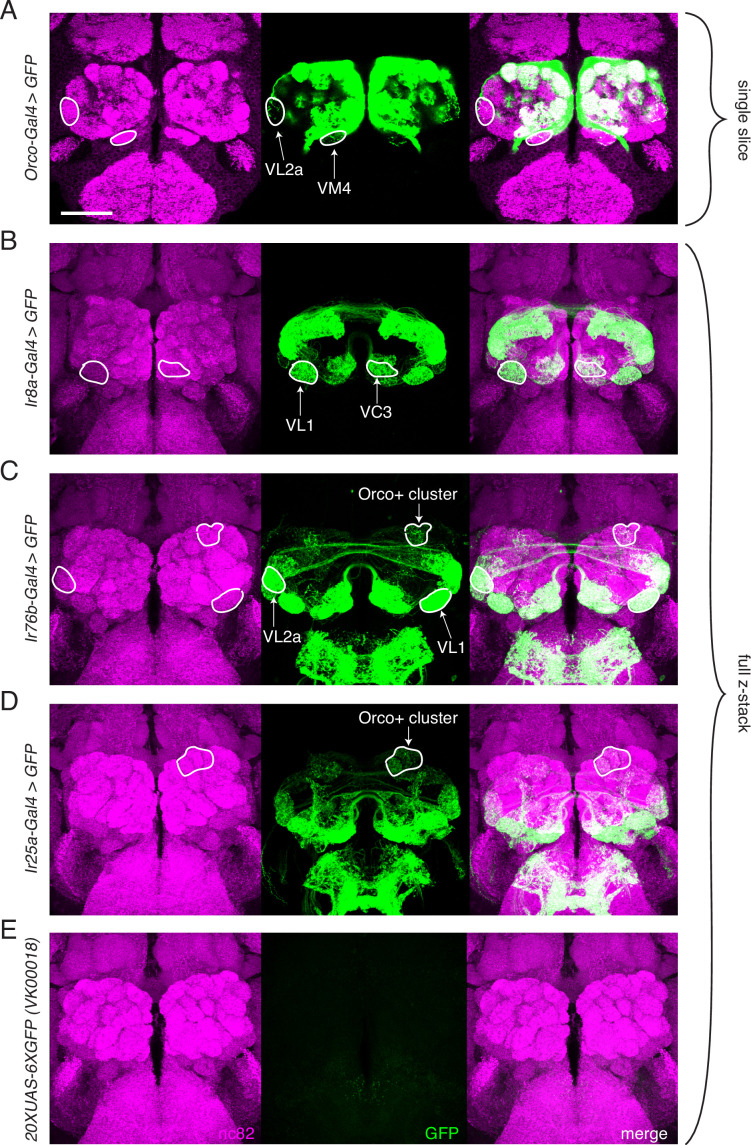
Figure 3—figure supplement 2. Transgenic co-receptor Gal4 lines do not fully recapitulate knock-in expression.

Figure 3—figure supplement 2.

(A–E) Transgenic co-receptor Gal4 lines crossed to a strong UAS reporter reveal expression in some, but not all, of the additional glomeruli labeled by the respective knock-ins. Some discrepancies between the Gal4 and knock-in lines are enumerated here, but do not represent a comprehensive list. (A) Orco-Gal4 drives weak GFP expression in VL2a and VM4 glomeruli (outlined), but lacks expression in DL2, V, and VL1 glomeruli, which are labeled by the knock-in. (B) Ir8a-Gal4>GFP labels VC3 and VL1 (outlined), but lacks expression in the VM4 and VA3 glomeruli, which are labeled in the knock-in. (C) Ir76b-Gal4 labels the VL1 and VL2a glomeruli (outlined), but lacks expression in the VA5 and DC3 glomeruli that are consistently labeled in the knock-in. Additionally, it drives GFP expression in Orco+ glomeruli not seen in the knock-in (outlined). (D) Ir25a-Gal4 labels some Orco+ glomeruli (outline includes DA2, DA3, DA4m, and DA4l), but is lacking expression in many glomeruli labeled by the knock-in. (E) The UAS-GFP control line has weak leaky expression in the subesophageal zone (SEZ) but no expression in the antennal lobe (AL). (A) shows single slice to visualize weakly labeled glomeruli; (B–E) are maximum intensity projections of z-stacks. Scale bar = 50 µm. See also Figure 3—source data 1.