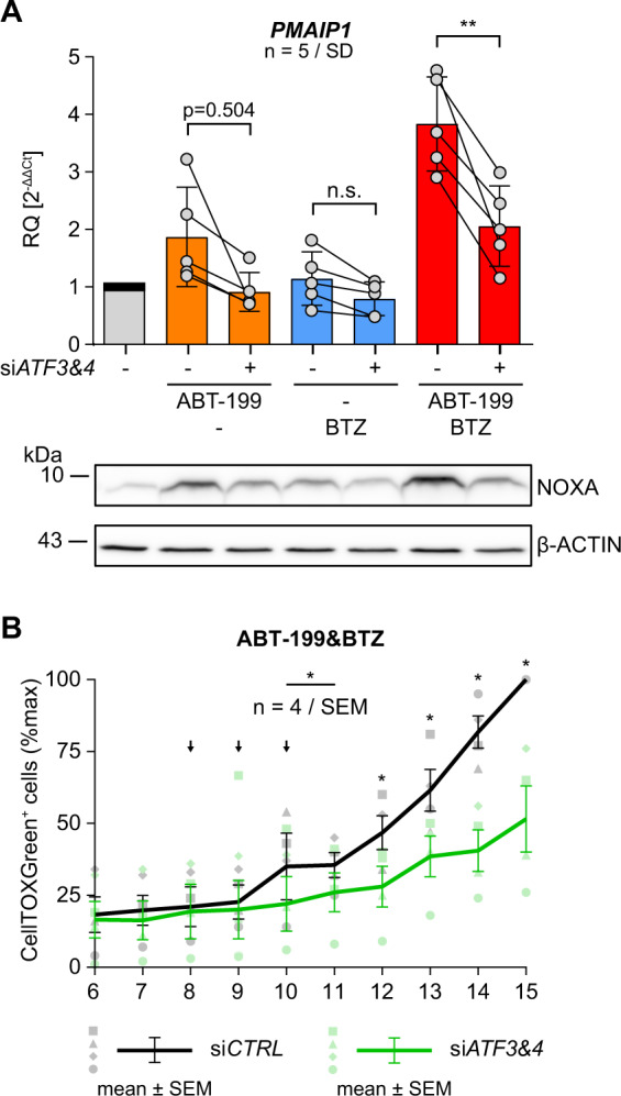
Fig. 5. Knock-down of ATF3&ATF4 reduces ABT-199&PI induced PMAIP1/NOXA expression resulting in diminished cell death.

A SW982/WT cells were transfected with siCTRL or siATF3/siATF4 and incubated with 15 µM ABT199, 5 nM BTZ or both for 8 h (+ 10 µM Q-VD-OPh). Aliquots from identical samples were analyzed by qRT-PCR and Western blot for expression of PMAIP1/NOXA (β-ACTIN as loading control). Graphs show mean values and individual data points. Statistical significance was analyzed by an unpaired student´s t test. B SW982/WT cells transfected with siCTRL or siATF3/siATF4 were incubated with 15 µM ABT-199 in combination with 5 nM BTZ. Medium was supplemented with CellTOX Green and cells were monitored by fluorescence microscopy imaging at the indicated time points. Graph shows the number of CellTOX Green positive cells as mean values ± SEM from 4 independent experiments. Statistical significance was calculated by a paired student´s t test. Arrows indicate excluded time points 8–10 h (■/□). BTZ bortezomib, CTRL control.
