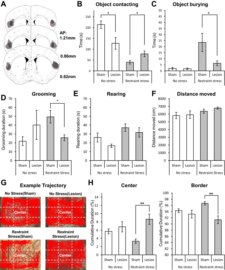
Figure 3.
Electrolytic lesion of the aIC in fear and anxiety-like behaviors. (A) Histological evaluation of the extent of damage made with electrolytic lesions in a series of sections of the insular cortex. Darker areas indicate complete ablation. Lighter areas indicate partial tissue damage. (B) Duration of contacting a restrainer-resembling object in four different groups: no stress (sham) n = 8, no stress (lesion) n = 7, restraint stress (sham) n = 9, and restraint stress (lesion) n = 8. (C) Burying of a restrainer-resembling object in different groups (same groups as in B). (D) Cumulative grooming duration difference between groups. (E) Cumulative duration of rearing behavior of different groups. (F) Total distance moved in each group (same group in B) during the experiment. (G) Example trajectory of four different groups. Red lines indicate trajectory of a mouse during the test. White dashed lines show how the center and border zones are defined in each experimental cage. (H) Cumulative duration in the center or border zone of different groups. (B–F, H) All data are presented as mean ± SEM. Mann–Whitney U test was used to assess statistical difference between groups within the no-stress and the restraint-stress group (*p < 0.05, **p < 0.01).

