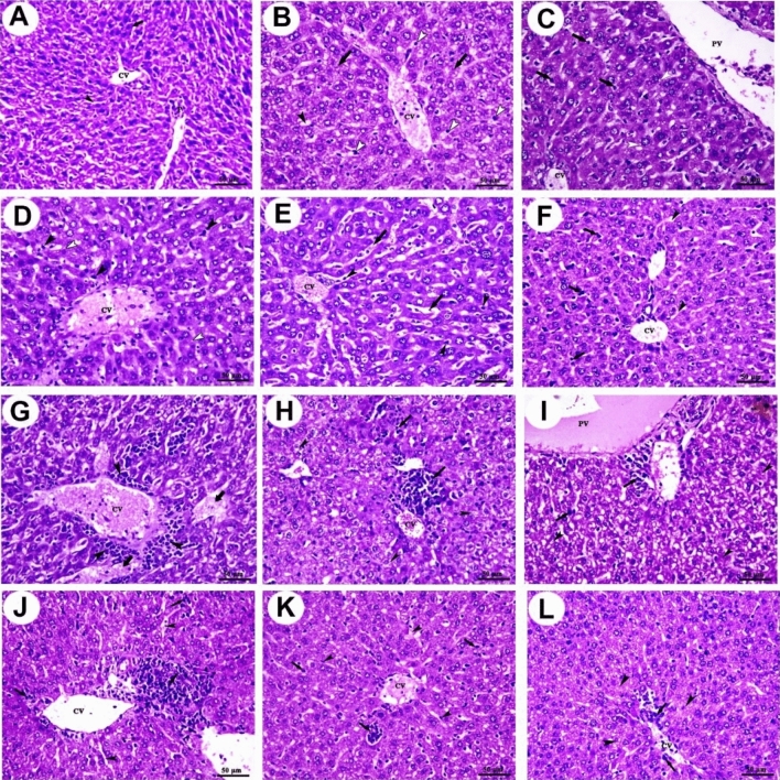
Figure 3.
Photomicrographs of mice liver sections stained with H&E. (A) Control group shows central vein (CV), portal vein area (P), polyhedral-shaped hepatocytes (arrow), and blood sinusoids (arrowhead). (B) Amy group shows congested central vein (CV), mild dilation of blood sinusoids (black arrowhead), mild degeneration of hepatocytes (arrows), and mild Kupffer cells activity. (C) SorIP group shows congested central vein (CV), portal vein (PV), mild vacuolation of hepatocytes (white arrowheads), and moderate Kupffer cells activity (arrows). (D) SorOS group shows congested central vein (CV), mild vacuolation of hepatocytes (white arrowheads), and moderate Kupffer cells activity (black arrowheads). (E) Amy + SorIP group shows congested central vein (CV), congestion, and dilation of blood sinusoids (arrows) in addition to Kupffer cells activity (arrowheads). (F) Amy + SorOS group shows mild congestion of central vein (CV), mild dilation of blood sinusoids (arrowheads), and a moderate increase of Kupffer cells activity (arrows). (G) EAC group shows aggregations of pleomorphic, hyperchromatic, and darkly basophilic cells (arrowheads) around central vein (CV) and hepatocellular necrosis (arrows). (H) EAC + Amy group shows a moderate number of pleomorphic cells (arrows) with moderate degeneration (arrowheads). (I) EAC + SorIP group shows a small focal area of pleomorphic cells (arrows) with moderate degenerative changes (arrowheads). (J) EAC + SorOS group shows a moderate focal area of pleomorphic cells (arrows) and mild dilation of some blood sinusoids (arrowheads). (K) EAC + SorOS group shows the smallest focal area of the pleomorphic cells (arrows) with the mildest congestion in some blood sinusoids (arrowheads). (L) EAC + Amy + SorOS group shows small diffuse pleomorphic cells (arrows) with mild vacuolar degeneration of hepatocytes (arrowheads). Scale bars = 50 µm.

