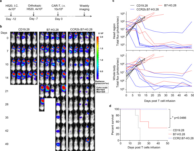
Fig. 6. CCR2b co-expressing B7-H3.CAR-T cells show superior antitumor activity in the H520 lung orthotopic and brain co-xenograft tumor model.
a Schematic of the H520 lung orthotopic and brain co-xenograft tumor model in NSG mice. Briefly, the FFluc-H520 (4 × 10⁴) cells were implanted into the right hemisphere of the brain of NSG mice by i.c. injection. Five days later, 4 × 10⁵ FFluc-H520 cells were implanted into the left lung by intrathoracic injection. Seven days later, mice were treated with CD19.28, B7-H3.28, or CCR2b.B7-H3.28 CAR-T cells (10 × 106 cells/mouse) by i.v. injection. b Representative tumor BLI images. Days represent the days post T-cell infusion. c The BLI kinetics of the FFluc-H520 tumor growth in the model shown in (b), BLI signals from the brain (top) and whole body (bottom) are illustrated, respectively (n = 5 mice/group). d Kaplan–Meier survival curves of mice in (b), n = 5 mice/group, *p = 0.0486 for B7-H3.28 vs. CCR2b.B7-H3.28, *p = 0.0181 for CD19.28 vs. B7-H3.28, **p = 0.0026 for CD19.28 vs. CCR2b.B7-H3.28, χ2 test. Source data for (c, d) are provided as a Source Data file.

