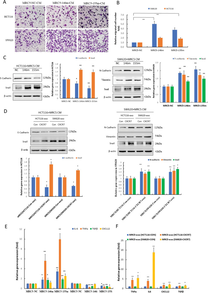
Fig. 6. CAF activation reciprocally promoted invasion and metastasis of CRC.
A, B Invasive capacity of CRC cells (HCT116 and SW620) co-cultured with CM from MRC-5 cells transfected with miR-146a-5p and miR-155-5p mimics (146m, 155m) was determined by transwell assay and statistical analysis was performed. **p < 0.01 vs. negative control (NC). C Western blot analysis of the expression levels of EMT-related proteins in HCT116 and SW620 cells treated with CM from MRC-5 cells transfected with miR-146a-5p and miR-155-5p mimics (146m, 155m). D Western blot analysis of the expression levels of EMT-related proteins in HCT116 and SW620 cells treated with CM from MRC-5 cells incubated with exosomes derived from CRC cells overexpressing CXCR7 and control. Densitometry and statistical analysis were performed. *p < 0.05, **p < 0.01 vs. control. E RT-qPCR analysis of IL-6, TNFα, TGFβ, and CXCL12 mRNA levels in MRC-5 cells transfected with miR-146a-5p and miR-155-5p mimics (146m, 155m) and inhibitors (146i, 155i). *p < 0.05, **p < 0.01 vs. negative control (NC). F RT-qPCR analysis of the mRNA levels of cytokines TNFα, IL-6, CXCL12, and TGFβ in MRC-5 cells treated with exosomes derived from HCT116CXCR7 and SW620CXCR7 cells and the corresponding controls. *p < 0.05, **p < 0.01 vs. control.

