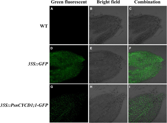
FIGURE 1.
Subcellular location of green fluorescent protein (GFP) and PsnCYCD1;1-GFP protein in tobacco root tip. Green fluorescent (A,D,G): green fluorescence signal; bright field (B,E,H): white light; and combination (C,F,I): combined signals of different fluorescence. WT, wild-type; 35::GFP, pROKII-GFP, empty vector control; 35S::PsnCYCD1;1-GFP, fusion vector of pROKII-PsnCYCD1;1-GFP.

