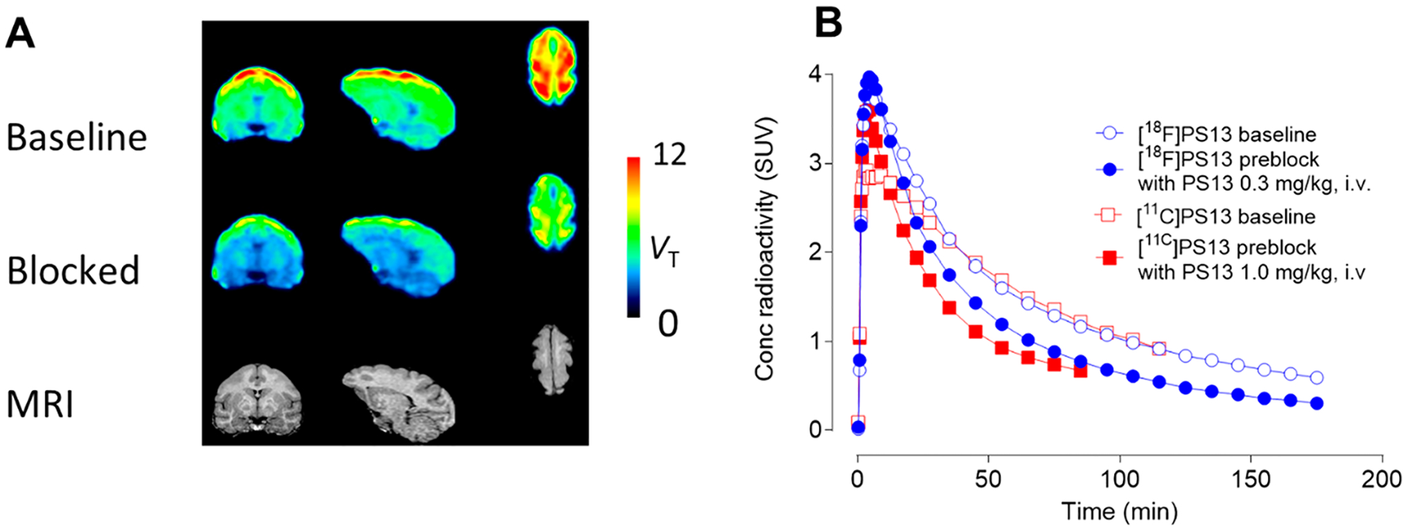
Figure 6.

PET studies of brain in monkey A with [18F]PS13 (carrier PS13:6.6 nmol/kg at baseline). Panel A: regional VT images from baseline scan (top row) and blocked scans (with PS13, 0.3 mg/kg, i.v.) (middle row), and corresponding MRI images (bottom row). Left column: coronal images; middle column: sagittal images; right column: horizontal images. Highest radioactivity uptake occurred in the prefrontal and parietal cortices. Panel B: whole brain time-activity curves at baseline and after preblock obtained with [18F]PS13. Corresponding curves obtained in a different monkey with [11C]PS13 from the study in ref 17 with a much lower PS13 carrier dose (0.19 nmol/kg at baseline) are shown for comparison.
