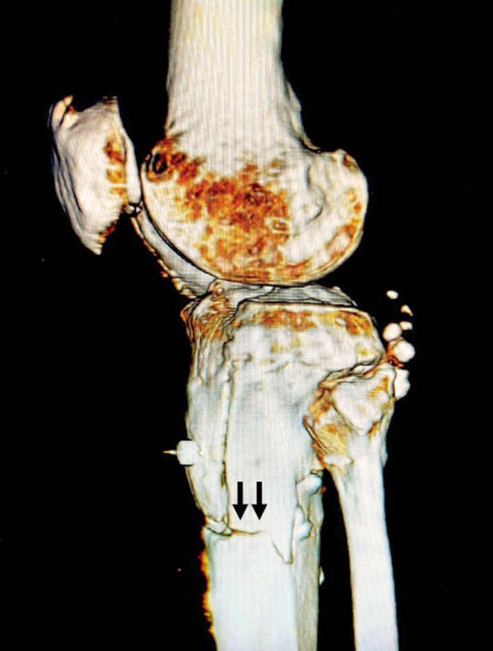
FIGURE 4.

Three‐dimensional computed tomography image after open‐wedge distal tuberosity osteotomy immediately prior to the second surgery. An unexpected fracture from the lateral distal edge of the descending osteotomy to the distal second screw hole was observed (arrows)
