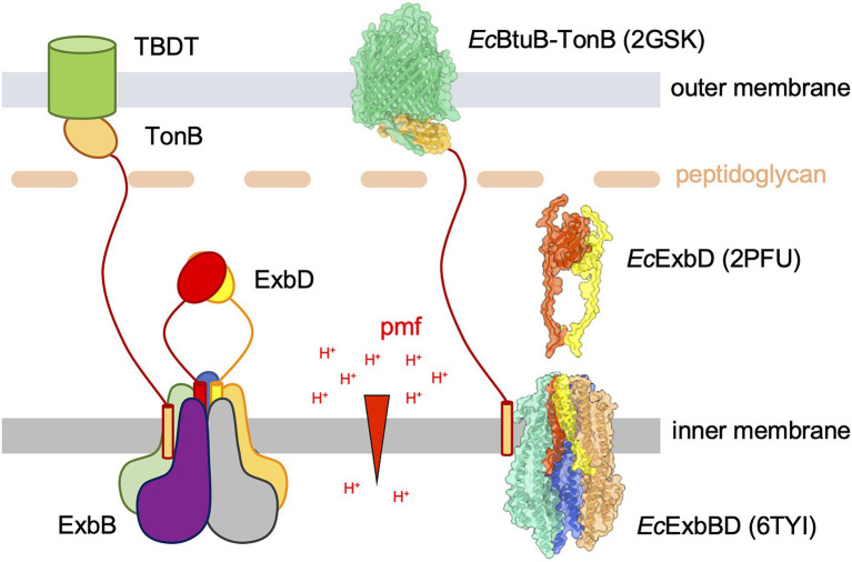
Figure 1.
A schematic representation of the Ton uptake system (left) and molecular models of the different components (right). Left panel: the TonB-dependent transporter (TBDT, green cylinder) is anchored in the outer membrane (OM). The ligand binds on the extracellular face of the TBDT and exposes a conserved domain called the TonB box to the periplasmic side. The TonB–ExbB–ExbD (TBD) complex is anchored in the inner membrane (IM) and uses the proton motive force (pmf, proton gradient across the IM, symbolized with the red arrowhead) to generate force and movement. The TBD complex is made of a pentamer of the ExbB subunit (blue, orange, grey, purple, and green) that defines a central pore in which a dimer of ExbD subunits (red and yellow) resides. The TonB subunit (gold) binds at the periphery of the ExbBD subcomplex. The elongated periplasmic domain of TonB allows its C-terminal globular domain to reach the OM and form a stable interaction with the TBDT TonB box. It is hypothesized that the ExbBD subcomplex forms the proton channel and that the energy derived from proton translocation is propagated through the TonB subunit to the TBDT, eventually opening a channel into the TBDT and allowing the bound ligand to diffuse into the periplasm. Right panel: molecular representations of known components of the Escherichia coli Ton system. The structural models are shown with ribbons and molecular surfaces. The color coding is the same than for the schematic view on the left. The TM and flexible periplasmic domain on the EcTonB subunit are not known and shown as in the schematic representation. The crystallographic structure of EcBtuB (green) in complex with the EcTonB periplasmic domain (gold) is represented in the OM (pdb 2GSK; Shultis et al., 2006). Two models of the NMR structure of the EcExbD periplasmic domains are shown in red and yellow (pdb 2PFU; Garcia-Herrero et al., 2007). The cryo-EM structure of the EcExbBD complex is shown in the IM (pdb 6TYI; Celia et al., 2019). The grey and purple ExbB subunits are not represented in order to show the ExbD TM domains. Molecular graphics have been performed with UCSF ChimeraX (Pettersen et al., 2021).

