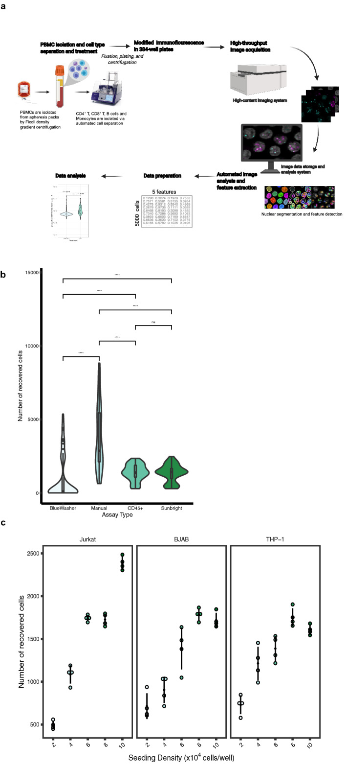
Figure 1.

HiIDDD: High-throughput imaging of DNA damage in human immune cells. (a) Schematic representation of the high-throughput imaging methodology to detect DNA damage in CD4 + T, CD8 + T, B cells and monocytes. The pipeline involves a modified immunofluorescence assay, imaging using an automated high-content screening system, image analysis for segmentation and fluorescence intensity cellular features extraction, and statistical analysis. Created with BioRender.com. (b) Determination of seeding efficiency using DAPI staining ~ 15 h after seeding. Additional substrates were added to poly-D-lysine plates; cells were seeded using a BlueWasher liquid handler unless indicated otherwise. Values represent at least 48 replicate wells per condition; box represents first and third quartiles. (c) Jurkat, BJAB, and THP-1 cells were seeded at densities ranging from 2–10 × 104 cells/well in a 384-well format. The total number of detected nuclei by DAPI is indicated. N = 4 wells per condition with at least 467 cells per condition.
