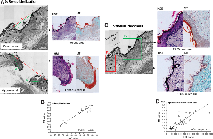
FIG. 3.
Re-epithelization and epithelial thickness index. Representative histology sections with examples on (A) Assessing the distance of axis covered by new epithelium (a and b – green line) and the distance of the minor axis between the original wound edges (c – red dotted line), in either a closed wound or open wound. (B) Spearman's correlation comparing re-epithelization assessed in paired MT an H&E sections. (C) Assessing epithelial thickness in the wound area (P2) and uninjured healthy skin (P1) by taking five measures (white dotted lines) in each area. (D) Spearman's correlation comparing epithelial thickness index as assessed in paired MT an H&E sections. Color images are available online.

