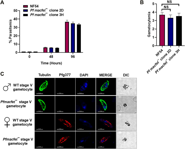
FIGURE 4.
Pfmacfet¯ parasites grow normally as asexual parasites and undergo gametocytogenesis. (A) Pfmacfet¯ (clone 2D and 3H) parasites grew at a similar rate as WT PfNF54. Data were averaged from three biological replicates and presented as the mean ± standard deviation (SD). (B) Bar graph shows gametocytemia for WT PfNF54 and Pfmacfet¯ parasites (clone 2D and 3H) measured on day 15. Data were averaged from three biological replicates and presented as the mean ± standard deviation (SD). (C) IFAs performed on thin blood culture smears for WT PfNF54 or Pfmacfet¯ (clone 3H) using α-tubulin (green), a male-specific marker, and Pfg377 (green), a marker for female gametocytes. Representative images are shown. DIC, differential interference contrast. DAPI, 4′,6-diamidino-2-phenylindole, Scale bar = 5 μm.

