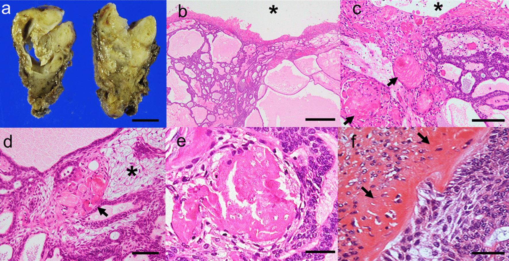
Fig. 3.

Macroscopical and histological findings. a The surgical specimen was a tan-white, lobulated solid cystic mass. b, c The cystic space was lined with a thick wall containing polygonal epithelial cells and ghost cells (arrows), similar to a calcifying odontogenic cyst, and translated the cribriform or plexiform basaloid component (asterisk, cystic space; hematoxylin and eosin [HE] staining). d The tumour nest with satellate reticulum-like cells including ghost cells and peripheral palisading (asterisk, satellate reticulum like nest; arrows, ghost cells; HE staining). e Ghost cells with prominent eosinophilic homogenisation and an absence of nuclei (HE staining). f Dentinoid material deposition adjacent to the tumour nest (arrows, dentinoid material; HE staining). Scale bar = 1 cm (a), 500 µm (b), 200 µm (c), 100 µm (d), and 50 µm (e, f). Images were taken using an OLYMPUA BX53 and scaled with PLYMPUS cellSens Standard
