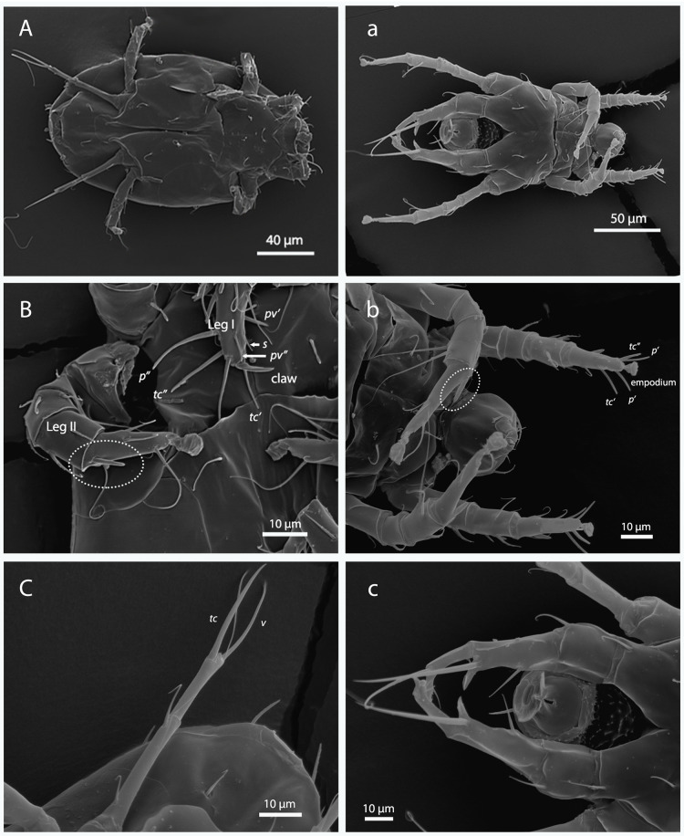
Fig 2.
Morphology of female (A-C) and male (a-c) Polyphagotarsonemus latus. A-a) ventral panoramic view; B-b) chaetotaxy of tibiotarsus of Leg I [eupathidium p´ (anteroproral), p” (posteroproral), tc´ (anterotectal), and tc” (posterotectal); setae pv´ (anteroprimiventral), pv” (posteroprimiventral) and s (subunguinal)] and solenidion of Leg II (within the ellipse); C-c) Leg IV [setae v" (posteroventral) and tc" (posterotectal)].

